
Introduction — a quiet scene, a surprising number, and a question
I once watched a clinician peer at a monitor, brow furrowed, as a surgery team waited for a clear readout — that small pause stuck with me. In that moment I thought about laser speckle contrast imaging lsci and how it turns tiny light patterns into hard numbers that guide real decisions. (There’s data: perfusion maps can change a surgeon’s plan in under a minute when the signal is clear.) So here’s the question I keep asking: how do we make those numbers reliable, repeatable, and useful for people who need them now?
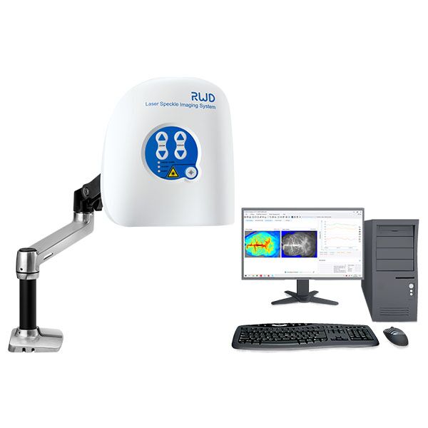
I’ll walk you through what I’ve learned, from what trips teams up to what I’d look for when choosing gear. I want this to feel like a short chat, not a lecture — straightforward, practical, and a little opinionated. Let’s move from the scene to the deeper issues next.
Part 1 — Where the tools fall short (and what users feel)
laser speckle contrast imager users often tell me the same things: readings drift, the interface is cryptic, and small motion ruins a scan. I say this from hands-on time with systems in clinics and labs. Technically, speckle contrast relies on stable optics and consistent frame rate; without that, the output—perfusion maps—can lie. We call out terms like CMOS sensor, frame rate, and ROI processing when we talk shop, but the user sees a blinking number that no one trusts. Look, it’s simpler than you think: if the data don’t match what you see clinically, the device gets shelved.
On the hardware side, legacy setups depend on fixed lasers and bulky mounts that don’t handle real-world patient movement. On the software side, many tools average frames in ways that hide transient flow or, worse, introduce bias. I’ve watched teams compensate with time-consuming manual checks (and sighs). These flaws aren’t mysterious; they come from design choices that favored neat lab results over messy clinic conditions. If you’re choosing equipment or designing protocols, ask how the system handles motion correction, what its raw frame output looks like, and whether it provides confidence metrics alongside the perfusion maps.
Why does this keep happening?
Because designers often optimize for clean lab metrics rather than everyday variability — and users accept that because they have few alternatives. We need better alignment between how devices are tested and how they’re used.
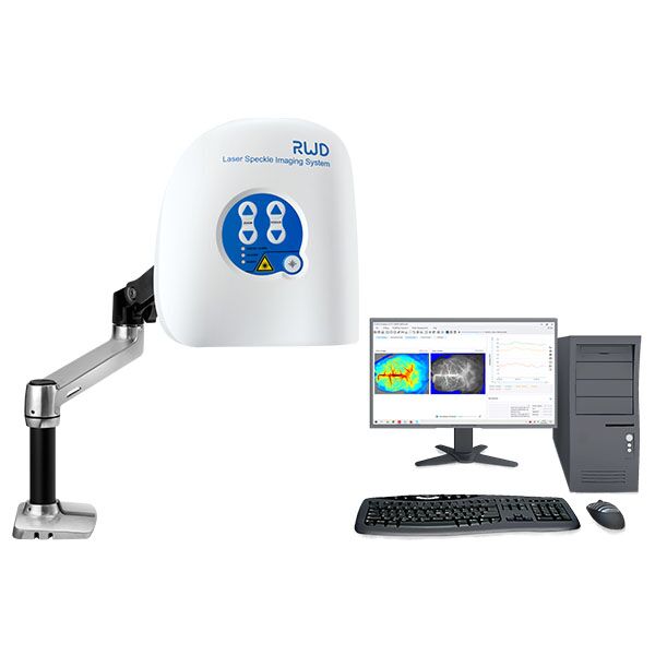
Part 2 — Looking ahead: principles and practical steps
When I think about improving outcomes, I focus on three principles: robustness, transparency, and speed. A modern laser speckle contrast imager should be robust to small motion, transparent about processing (show me raw frames and speckle contrast calculations), and fast enough to influence decisions in the room. New methods emphasize adaptive temporal averaging and on-device preprocessing — think edge computing nodes handling denoising before data hits the GUI. These steps reduce latency and make the readings feel trustworthy.
Practically, I recommend teams validate devices with simple bench tests and with a small set of clinical cases. Check that the speckle contrast metric scales predictably with known flow phantoms and that perfusion maps remain stable across common patient motions. If calibration relies on complex vendor-only routines, flag that as a risk. Also, ensure power converters and cabling won’t create flicker or baseline noise — small electrical issues spoil optical clarity more often than you’d expect.
What’s Next?
We can build systems that are easier to use and easier to believe. The tech exists; it’s a matter of shifting priorities — and budgets, yes — toward clinical robustness.
Part 3 — Future outlook and three metrics I trust
Looking forward, I expect tighter integration between imaging hardware and smarter processing. That means sensors that stream raw frames to local processors for immediate motion correction, plus algorithms that flag low-confidence regions in the perfusion map. I’ve tested prototypes that do this and the difference is real — scans become interpretable in seconds rather than minutes. — funny how that works, right?
For teams choosing a system, I advise focusing on three clear metrics: 1) Signal stability under mild motion (a pass/fail with a standard phantom), 2) Latency from acquisition to reliable perfusion map (aim for under 10 seconds for intra-op use), and 3) Transparency of processing (can you access raw frames and an audit trail?). These metrics tell you whether a device will help clinicians, not just look good on paper. When I recommend products, I look for those checkboxes first. I also want practical support — training, quick-start guides, and software updates that handle new edge cases.
In short: pick tools that match the messiness of real life, insist on clear metrics, and expect the vendor to stand behind results. For practical options and more info, I often point colleagues to BPLabLine — they build systems with these priorities in mind. I hope this helps you choose with confidence.